A staff member’s participation in a voluntary research study detected a sight-threatening disease which was misdiagnosed by opticians.
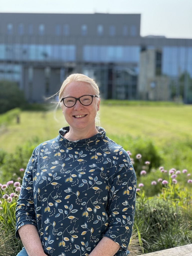
Tammie Farley, College of Science Events and Schools Liaison Manager at the University of Lincoln, UK responded to an open call to take part in a study where her eyes were to be routinely photographed. The study was being ran by Dr Bashir Al-Diri, Associate Professor and Programme Leader at Lincoln School of Computer Science as part of his project titled ARIAL (Automated Retinal Image Analysis Lab).
She volunteered as someone with healthy eyes to provide Dr Bashir with photographs to help build software designed to automatically analyse retinal images. The images can detect vascular segmentation which, if untreated, can cause blindness.
In February 2022, an irregularity appeared on the photographs of Tammie’s eyes. A month prior, she had noticed that straight lines appeared to have kinks in them. Spreadsheets, for example, became especially difficult for her to see clearly. An optician had diagnosed age-related macular degeneration (AMD), though Dr Bashir believed that this was a misdiagnosis.
Through corresponding with a fellow researcher at the University of Sunderland, Dr Maged Habib, a Consultant Ophthalmology and Retina Specialist, Dr Bashir was able to confirm that Tammie had Central Serous Chorioretinopathy with CNV (choroidal neovascularisation) rather than AMD. This disease can be sight-threatening.
The first four months are crucial in determining whether a patient with this disease will lose sight or not. By taking part in the study, Tammie was correctly diagnosed quickly and able to receive treatment with Dr Maged in March this year at no charge.
“I am incredibly grateful to Dr Bashir for not only detecting the issue in my right eye but also his concern and continued monitoring of it over the past 15 months. Had Bashir not liaised with Dr Habib at Sunderland Eye Infirmary I would not have received such speedy treatment. If it had been left without treatment or undiagnosed, I could have been left with permanent central vision loss.”
Tammie Farley
Tammie’s experience is just one example of the exceptional work ARIAL is doing to protect people’s eyesight. Since the project began in May 2018, 12 per cent of the participants were found to have abnormal signs in their retinal images and were encouraged to follow up with their opticians and continue with the retinal scans to detect any further changes.
A further five per cent of the participants had started to develop more serious conditions that would have been undetected by the participant and were referred to their GPs for more investigations.
Dr Bashir Al-Diri, Lead Investigator of ARIAL, said:
“Our vascular system adapts to various conditions, age, and lifestyle activities. It is crucial to differentiate between changes due to pathology and those due to normal aging and lifestyle factors. However, this has been difficult in the past due to a lack of images from non-pathological participants.
Dr Bashir Al-Diri
“The ARIAL project aims to address this issue by collecting images over time to discover patterns and learn cause-and-effect relationships. By analysing these images and correlating them with other clinical data, the project hopes to develop a set of integrated techniques that will influence routine clinical patient care in the years to come.”
Dr Bashir was the first ever PhD student at Lincoln’s School of Computer Science and has been researching this subject for almost 20 years. To develop an artificial intelligence system that monitors changes to a patient’s eye, he requires considerably more photographs of eyes where any segmentation is manually measured and logged.
This manual process, however, is incredibly time consuming. He hopes to employ the assistance of medical students or colleagues to continue progressing the project which could have a transformative impact on UK provision of optical care.
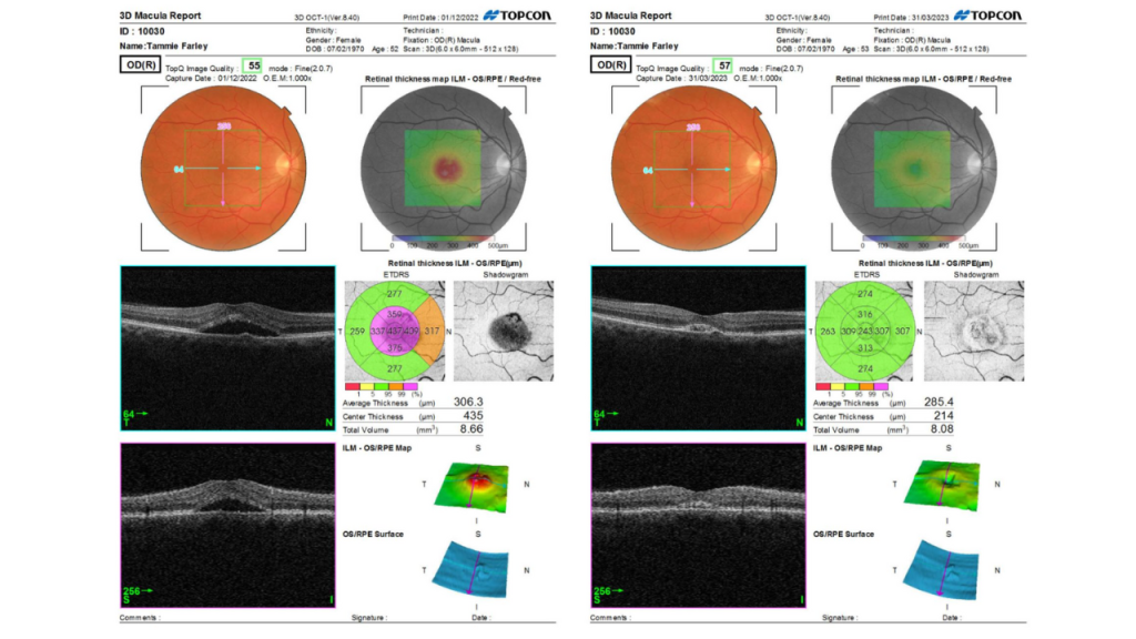
Image 1 shows the abnormality identified in Tammie’s eye in photos captured on 01/12/2022. Image 2 shows Tammie’s healthy eye report from 31/03/2023 following treatment. The retinal thickness shadowgraph indicates where the disease is present and shows the significantly increased thickness. A ‘hole’ is also visible on the left-hand photos which is shown to be repaired in Image 2. The fully green thickness graph displayed in Image 2 also shows the success of the treatment, with only faint traces of abnormality left on the shadowgraph.